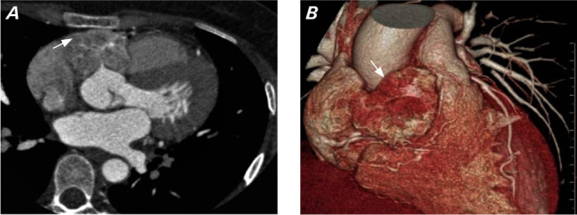
Fig. 1.

A) Multidetector computed tomography angiography image in axial view shows a lobulated heterogeneously enhancing mass (arrow) in the atrioventricular groove encasing ostia of the right coronary artery. B) Multidetector computed tomography angiography volume rendered image shows a mass in the right atrioventricular groove (arrow) extending superiorly between the aorta and pulmonary artery.
