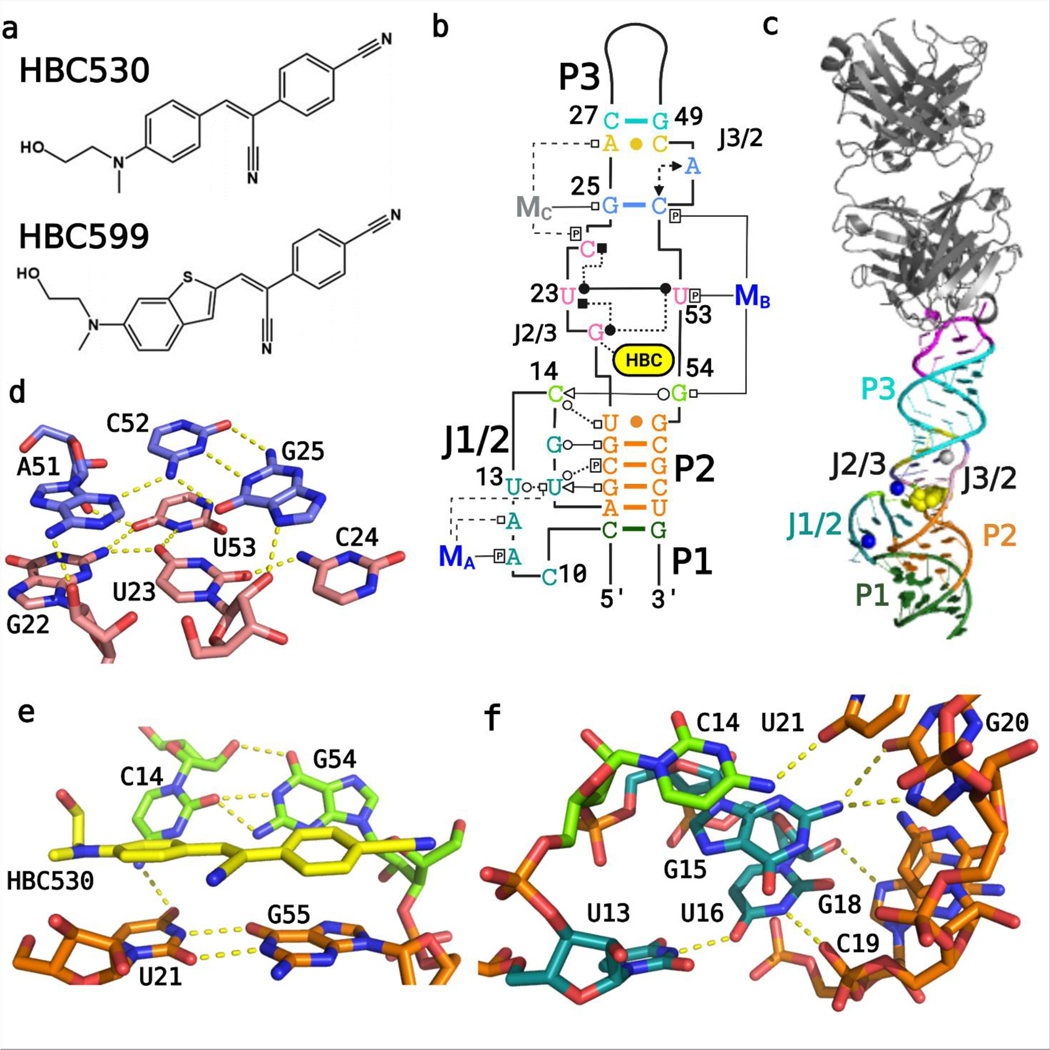
Figure 1.
Overall structure of the Pepper-BL3–6 aptamer. (A) Chemical structures of the HBC ligands, HBC530 and HBC599 activated by Pepper RNA. (B) Tertiary structure of the Pepper aptamer bound to HBC530 utilizing the Leontis−Westhof nomenclature for RNA-interaction classification,12 with color-coded regions, and the two Mg2+ cations that are visible in both structures (MA, MB; blue) and the Na+ ion visible in the HBC530 complex only (MC; gray). (C) Fab BL3–6 bound to Pepper with the HBC530 ligand. (D) Tetrad (pink) and triplex (blue) structures above the ligand from the HBC599 complex, without MC. (E) Tertiary interactions between C14 and G54, adjacent to the bound ligand HBC530. (F) Tertiary interactions between J1/2 and P2.

