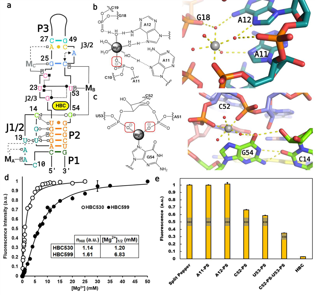
Figure 4.
Mg2+ sites identified in the crystallized Pepper-BL3–6 aptamers. (A) Tertiary structure diagram of Pepper bound to HBC530, with the magnesium ions (MA, MB) labeled. (B) Diagram and cartoon of the J1/2 Mg2+ (MA) with one inner-sphere contact to A11-PO. (C) Diagram and cartoon of the binding site Mg2+ (MB) with three inner-sphere contacts to G54−N7 and the C52 and U53 phosphates. The G54−C14 interaction is also highlighted. (D) 2 M NaCl-background fluorescence isotherm showing how fluorescence of Pepper-BL3–6 changes over [Mg2+], with nHill =1.14 ± 0.10 for binding HBC530 and nHill = 1.61 ± 0.09 for binding HBC599. With the saturated ion atmosphere from 2 M NaCl, Mg2+ binding is limited to non-monovalent binding sites on the aptamer. (E) Fluorescence of the split-Pepper system with phosphorothioate modifications to probe Mg−O inner-sphere interactions. 50 and 75% signals marked with dotted lines to gauge significance.

