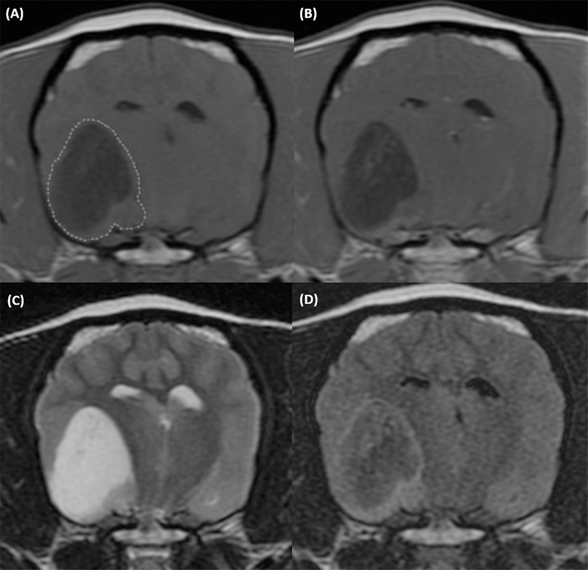
Figure 1.

T1-weighted precontrast (A), T1-weighted postcontrast (B), T2-weighted (C), and T2 FLAIR (D) transverse MR images of the brain of a 9-year-old female intact Boston Terrier with a grade 2 (low grade) oligodendroglioma. An ROI outlines the lesion on the T1-weighted precontrast image (dotted line). Acquisition parameters: transverse, slice thickness 3mm, slice spacing 3.3mm. T1W precontrast: FSE T1, TR 567ms, TE 11ms, NEX 4. T1W postcontrast: FSE T1, TR 567ms, TE 11ms, NEX 4. T2W: FSE T2, TR 3500ms, TE104ms, NEX 3. T2 FLAIR: IR, TR 8002ms, TE126ms, TI 2000ms, NEX 1.
