
Figure 2.
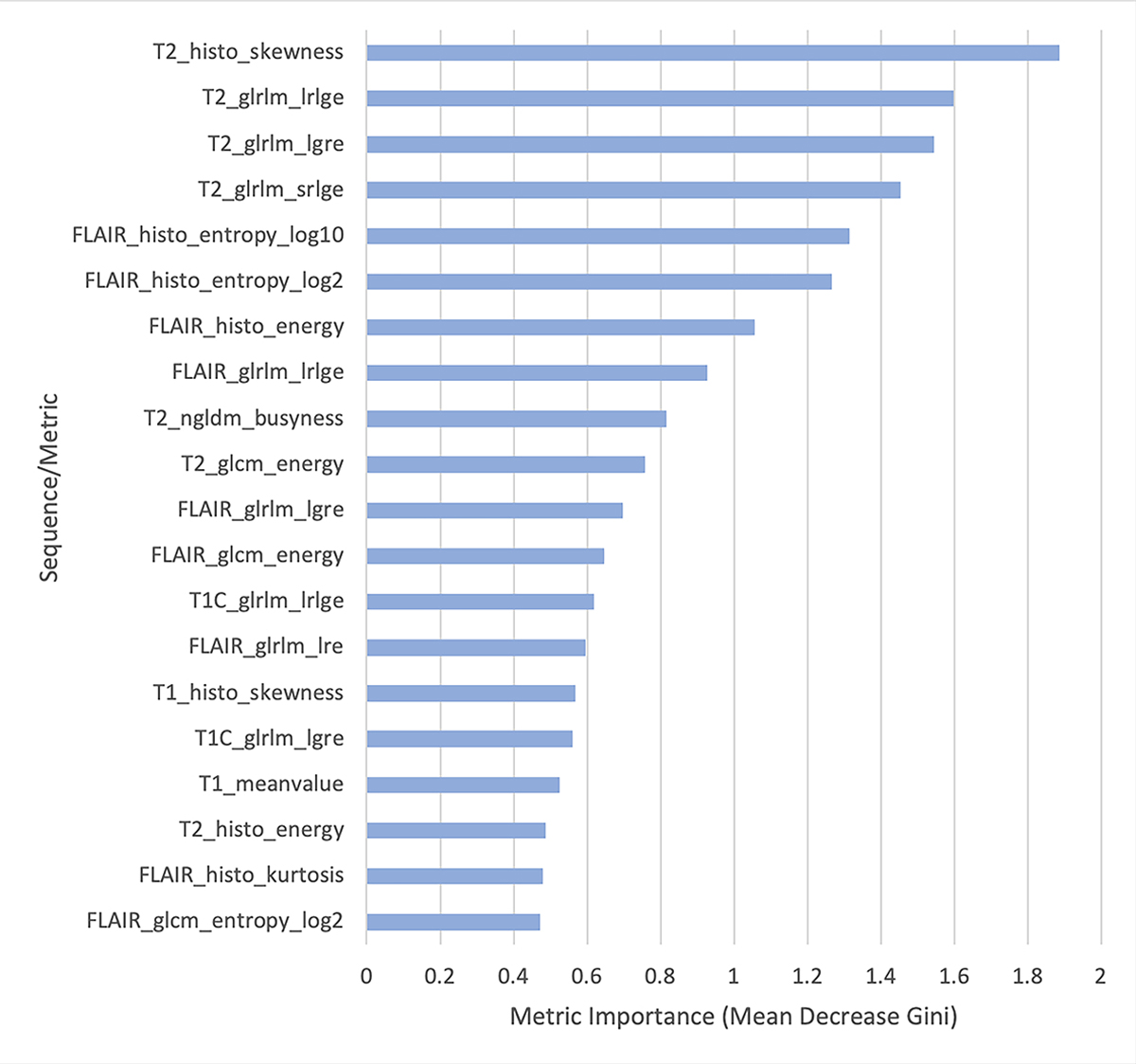
Variable importance plots from the random forest classifier differentiating inflammatory versus neoplastic brain disease in order of decreasing importance (top to bottom). This graph displays the 20 most discriminatory metrics in constructing the random forest classifier. histo, histogram; glrlm, grey-level run length matrix; ngldm, neighborhood grey-level different matrix; glcm, grey-level co-occurrence matrix; lrlge, long run low grey-level emphasis; lgre, low grey-level run emphasis; srlge, short run low grey-level emphasis; lre, long run emphasis. Magnetic resonance imaging (MR) sequences include T2 weighted (T2), fluid attenuated inversion recovery (FLAIR), T1 weighted (T1), and T1 plus intravenous contrast administration (T1C).