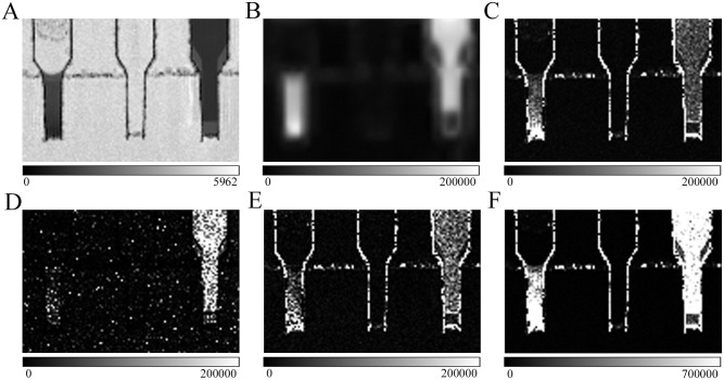
Fig 2. Comparison between several types of images for different phantoms.
(A) ADCb. (B) DKI. (C) ASM/A3. (D) ASM/S3. (E) PASM/A3. (F) ASM/A5. From left to right in every image, phantoms are high-cellularity bio-phantom, physiological saline phantom and 120 mM polyethylene glycol phantom. ADC, apparent diffusion coefficient; ADCb, ADC basic; DKI, diffusion kurtosis image; ASM, ADC subtraction method; ASM/A3, ASM division by ADCb three times; ASM/S3, ASM division by standard deviation image three times; PASM/A3, positive ASM division by ADCb three times; ASM/A5, ASM division by ADCb five times.

