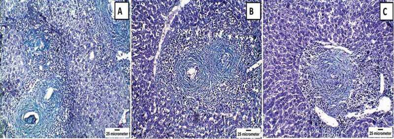
Figure 5.

Histopathological changes of Masson’s trichrome (MT)-stained livers of mice infected with Schistosoma mansoni twelve weeks post-infection showing (a) the control infected group with positive staining in the concentric fibrous layers surrounding the granuloma and in the wall of hepatic sinusoids (x200); (b) praziquantel-treated group with moderate positive staining (x200); and (c) mesenchymal stem cells-treated group with mild positive staining (x200). Note the reduction in the number and size of granulomas in both treated groups in comparison to the infected, untreated group.
