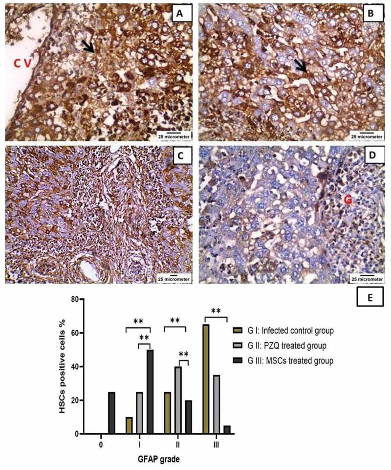
Figure 7.

Immunohistochemical staining of glial fibrillary acidic protein (GFAP) in the livers of mice infected with Schistosoma mansoni twelve weeks post-infection showing (a) & (b) the infected, untreated group with intensely immunostained hepatic stellate cells (HSCs) present in the walls of sinuses (black arrow) as well as in the perivenular areas (x400) CV = central vein; (c) praziquantel-treated group with moderate GFAP expression in HSCs in the sinusoidal wall (x200); (d) mesenchymal stem cells-treated group with mild GFAP expression (x400) G = granuloma; and (e) The percentage HSCs positive for GFAP expression in the liver of the studied experimental animal groups. ns: non-significant, * P values ˂ 0.05, ** P values ˂ 0.01, and *** P values ˂ 0.001.
