
Fig 7. Intralaminar circuit diagram:
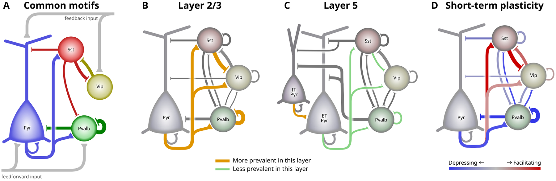
The cortical intralayer circuit differs across layer and with activity. A. Some commonly described elements of the intralaminar cortical circuit. Pvalb cells strongly inhibit pyramidal and other Pvalb cells, Sst cells provide broad inhibition, and Vip cells inhibit Sst cells to form a disinhibitory feedback pathway. B-C. Circuit diagrams showing connections between major subclasses in mouse L2/3 (B) and L5 (C). The width of connecting lines roughly represents connection probability and PSP amplitude. Connections that are prominent in each layer compared to the other are highlighted in orange, whereas green lines indicate connections that are less prevalent in that layer. For simplicity, connections between IT pyramidal and inhibitory in L5 (C) are omitted. D. Two complementary circuits that activate at different times. Red connections are facilitating and will be stronger during sustained activity. Blue connections are depressing and are strongest during quiescent periods.