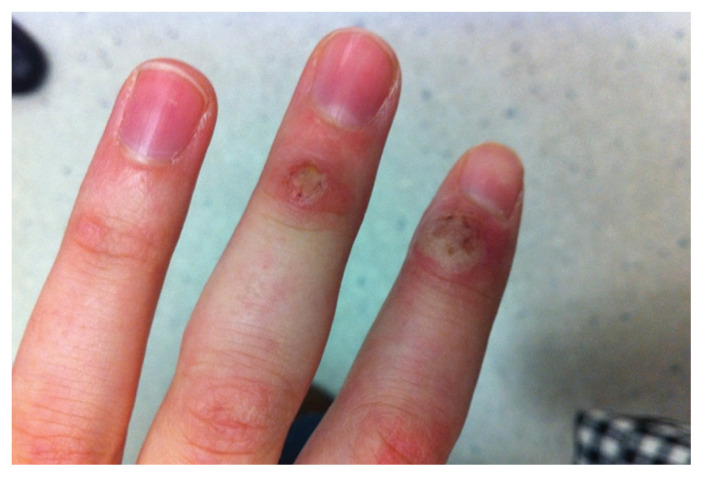
Figure 1:
Photograph of lesions on the left fingers of a 21-year-old woman with pseudocowpox. The lesion on her second finger had a slightly depressed ulcerative appearance; the lesion on her third finger was semi-firm and filled with fluid, in keeping with the exudative nodular phase. Both lesions contained purplish–red punctate areas and mild surrounding erythema.

