Figure 6.

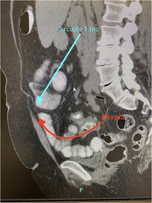
CT imaging demonstrates separation of the posterior sheath from the rectus abdominis at the arcuate line with herniated fat or viscus (sagittal imaging).

CT imaging demonstrates separation of the posterior sheath from the rectus abdominis at the arcuate line with herniated fat or viscus (sagittal imaging).