Fig. 6.
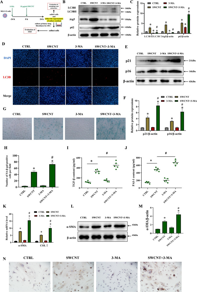
Inhibition of autophagy using 3-MA aggravated FMT in lung fibroblasts by promoting SWCNTs-triggered senescence in MLE-12 cells. MLE-12 cells were exposed to 10 μg/ml SWCNTs for 24 h with or without 3-MA pretreatment,, and then the cell-free culture medium, as CM, was used to culture primary lung fibroblasts for 48 h. A Scheme of workflow for evaluating the effects of 3-MA on autophagy and senescence in MLE-12 cells and FMT in lung fibroblasts after SWCNTs exposure in vitro. B, C Western Blotting analysis of Atg5, LC3BI, LC3BII and p62 protein expressions in MLE-12 cells (n = 3). D LC3B puncta in MLE-12 cells was observed by IF (200×). E, F Western Blotting analysis of p21 and p16 protein expressions in MLE-12 cells (n = 3). G SA-β-gal activity was determined by X-gal staining in MLE-12 cells (400×). H The number of SA-β-gal positive MLE-12 cells per field (n = 8). Contents of TGF-β (I) and PAI-1 (J) in the supernatants of culture medium quantified by ELISA (n = 5). K mRNA expressions of α-SMA and COLI in lung fibroblasts were measured by qRT-PCR (n = 4). L, M Western Blotting analysis of α-SMA protein expression in lung fibroblasts (n = 3). N COLI positive expression in lung fibroblasts was evaluated by IHC (n = 5) (200×). *P < 0.05 vs CTRL group. #P < 0.05 vs SWCNT group

