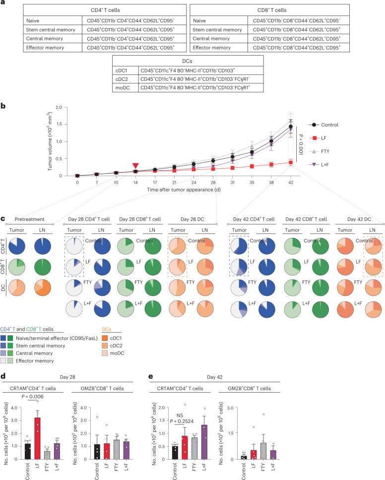
Fig. 2. Lymph node egress is necessary for l-fuc-triggered tumor suppression; l-fuc increases intratumoral CD4+ T stem and central memory cells.
a, Immune subpopulation markers use to profile by flow cytometry. b, Volumetric growth curves for SW1 tumors in C3H/HeN mice fed without (control, n = 3 mice) or with (l-fuc, n = 10 mice) l-fuc and treated with FTY720 (control mice were administered FTY720 (FTY) (n = 10 mice); l-fuc-supplemented mice were administered FTY720 (L + F) (n = 10 mice)). FTY720 was administered at 20 µg per mouse every 2 d starting on day 12, just before the initiation of l-fuc administration. c, Pie charts showing ratios of intratumoral or lymph node (LN)-resident CD4+ or CD8+ T cell subpopulations as well as DC subtypes from mice at days 14, 28 and 42 (each pie chart represents 4–5 mice). Assessment of cytotoxic CD4+ T cell populations (CRTAM+) and cytotoxic CD8+ T cell populations (GrzB+) from tumors at day 28 (d) (n = 5 mice for all groups except l-fuc, where n = 4 mice) and day 42 (e) (n = 5 mice each for control and l-fuc groups, n = 4 mice each for FTY and L + F groups). NS, not significant. Corresponding raw flow cytometric data for these charts are shown in Supplementary Table 1. The tumor growth curves and column charts show mean ± s.e.m. per group of mice.

