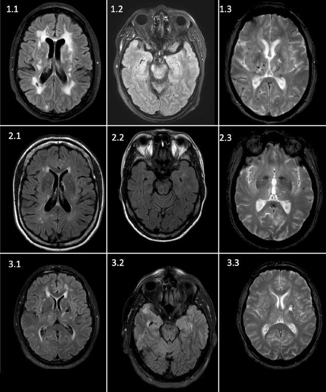
Fig. 1.
MRI images of the three CADASIL patients identified in our cohort. Patient 1 refers to the known CADASIL disease. 1.1: FLAIR sequence showing lacunar defects and extensive confluent white matter lesions, 1.2: FLAIR sequence, white matter lesions without emphasize on temporopolar region, 1.3: SWI sequence showing extensive microbleeds both thalamic and cortical. Patient 2 refers to the clinically suspected patient. 2.1: FLAIR sequence with extensive white matter lesions. 2.2: FLAIR Sequence, no emphasize on temporopolar, 2.3: T2* (“heme”) sequence showing supratentorial cortical microbleeds as well as thalamic microbleeds. Patient 3 refers to the clinically not suspected patient. 3.1 FLAIR sequence showing temporal white matter lesions and a lacunar defect in the thalamus, 3.2: FLAIR sequence, temporal white matter lesions, 3.3: T2* sequence, no microbleeds were shown

