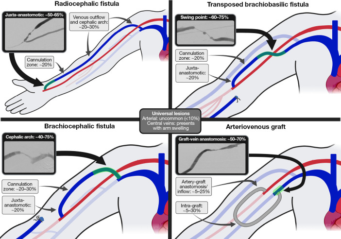
Figure 2.
Distribution and approximate frequency of stenotic lesions in different access configurations. The most common stenotic lesions are highlighted (green) with a representative image example: juxta-anastomotic stenosis in radiocephalic fistulae, CAS in brachiocephalic fistulae, swing point stenosis in transposed brachiobasilic fistulae, and graft-vein anastomotic stenosis in grafts. The frequency of different lesions are rough approximations determined from a conglomerate of sources, meant to provide an estimation of the relative occurrence of these lesions. The reader is referred to the source data in the cited articles for specific numerical data (8,10,12-15,19,34-48). CAS, cephalic arch stenosis.

