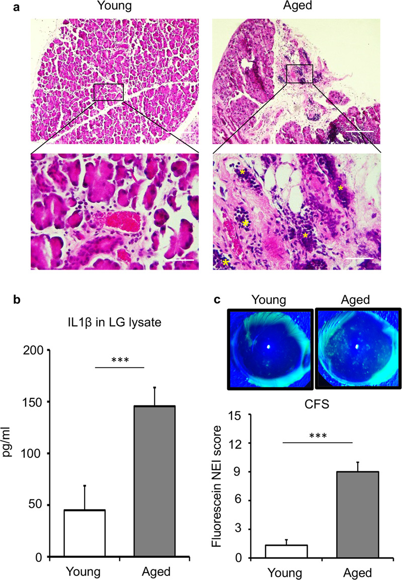
Fig. 3. Aged mice showing increased immune cell infiltration in the lacrimal gland and corneal epitheliopathy.

a Cross-sections of young and aged C57BL/6 mice lacrimal glands were stained with hematoxylin and eosin to visualize immune cell foci (yellow stars) and acinar atrophy (scale bar, 100 µm (upper); 20 µm (lower)). b Bar chart depicting protein levels of IL1β in the lacrimal gland lysates of young and aged mice, using ELISA analysis. c Representative slit-lamp images (upper panel) and cumulative bar chart (lower panel) measuring corneal fluorescein-staining (CFS) of young and aged mice. Cumulative data (mean ± SD) from three independent experiments are shown, with each experiment consisting of n of 4 to 6 mice/group. ***p < 0.001.
