FIGURE 1.
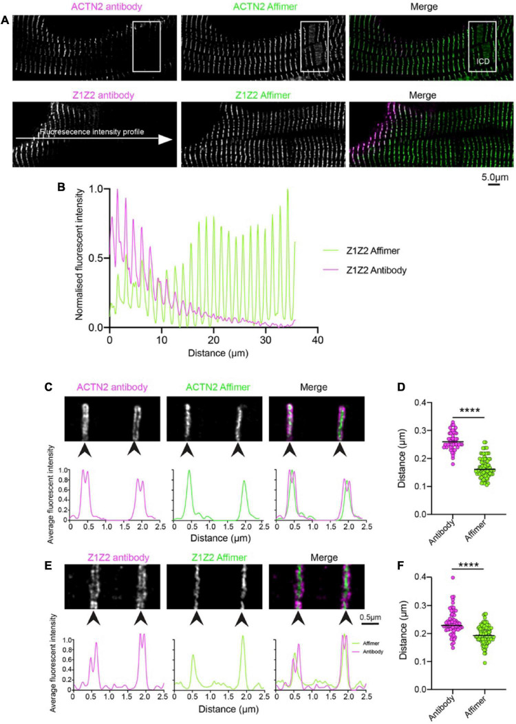
Comparison of staining heart sections using antibodies and Affimers to ACTN2 and titin Z1Z2 repeats. (A) Example confocal image of a region of a donor heart section stained using a primary antibody to ACTN2 or the Z1Z2 repeat of titin combined with a secondary fluorescent antibody, and with a dye-labelled Affimer. The boxed region shows the position of the ICD: intercalated disc. (B) Example of fluorescence intensity (normalized) for a line profile drawn across the cell for the ACTN2 antibody (magenta) and Affimer (green). Example 2D-STED images for Z-discs stained using the ACTN2 primary and secondary antibody combination and the dye-labelled ACTN2 Affimer (C) and the titin Z1Z2 antibody combination and Z1Z2 Affimer (E) are shown together with the associated profile plots for the labelling intensity across the Z-disc structures. (D,F) Measurements of the Z-disc widths for multiple Z-discs from sections labelled with the antibody combination and Affimers, using either the antibody images or the Affimer images. ****p < 0.0001.

