. 2023 Feb 9;24(3):224–234. doi: 10.3348/kjr.2022.0351

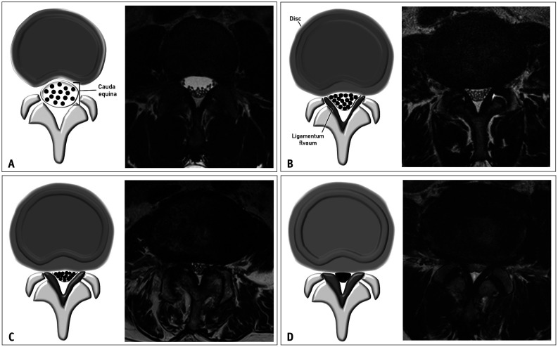
Fig. 1. The Lee grading system for lumbar central canal stenosis. A. Schematic drawing of normal central canal and the corresponding T2-weighted axial magnetic resonance image at the L3/4 disc level show no obliteration of the anterior cerebrospinal fluid space. B. Schematic drawing of mild central canal stenosis and the corresponding T2-weighted axial magnetic resonance image at the L4/5 disc level show mild obliteration of the anterior cerebrospinal fluid space and all cauda equina clearly separated from each other. C. Schematic drawing of moderate central canal stenosis and the corresponding T2-weighted axial magnetic resonance image at the L4/5 disc level shows moderate obliteration of the anterior cerebrospinal fluid space and some cauda equina aggregation. D. Schematic drawing of severe central canal stenosis and the corresponding T2-weighted axial magnetic resonance image at the L4/5 disc level show severe obliteration of the anterior cerebrospinal fluid space, marked compression of the dural sac, and the entire cauda equina appearing as single bundle.

Fig. 1