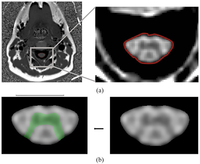
Figure 2.
Example of cervical spinal cord area imaging techniques. (a) Axial slice of PSIR C2-C3. Axial slice of a one-dimensional PSIR C2-C3 spinal cord image. The cord is segmented using automated method and the border is shown delineated here in red. (b) Gray matter segmentation. The spinal cord is cropped and up sampled 10 times on all dimensions. The gray matter segmentation is then performed on this processed image. The resulting segmentation is shown in green.
PSIR: phase-sensitive inversion recovery.

