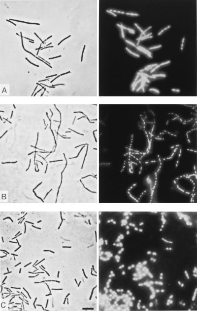
FIG. 6.
Same-field micrographs of B. cereus cells obtained by phase-contrast microscopy (left panels) and epifluorescence microscopy after DAPI staining (right panels). (A) Shortly after 0.05% polyP was added (1 h), beginning filamentation was apparent, but this process did not affect chromosome replication and coordinated nucleoid segregation in each individual elongated cell body. (B) Addition of 5 mM Ca2+ (after 3 h) reinitiated normal division events (septation) in the multinucleoid filaments. (C) Almost normal cell growth, division, and nucleoid distribution in the cells 3 h after Ca2+ was added. Bar = 10 μm.

