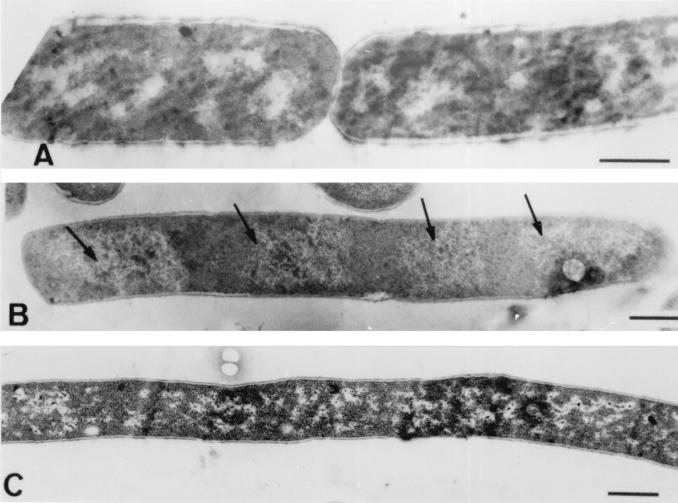
FIG. 7.
Transmission electron micrographs of thin sections of B. cereus WSBC 10030 cells following treatment with polyP. (A) Control cells before polyP addition. (B) Cells treated with 0.05% polyP after 30 min. The arrows indicate segregated nucleoids. (C) Cells treated with 0.05% polyP after 2 h. A complete lack of septum formation and the resulting filamentation is clearly evident. Bars = 0.5 μm.

