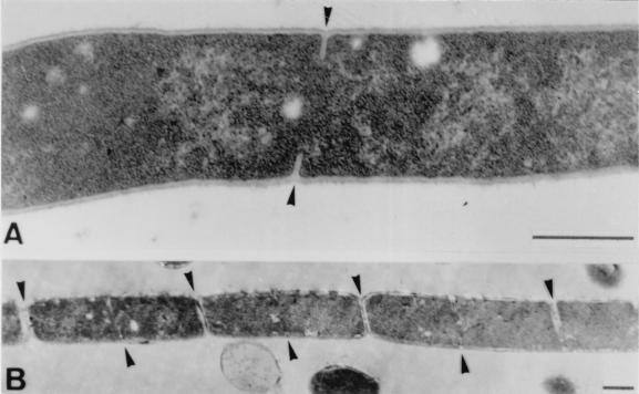
FIG. 8.
Transmission electron micrographs of thin sections of polyP-treated B. cereus cells (i.e., filaments) following addition of 5 mM Ca2+. (A) After 10 min, ring-shaped septum formation and growth resumed (arrowheads). (B) After 45 min, newly formed septa were completed, and cell division (i.e., separation of daughter cells) began (upper arrowheads), while a second cycle of septation is already evident (lower arrowheads). Bars = 0.5 μm.

