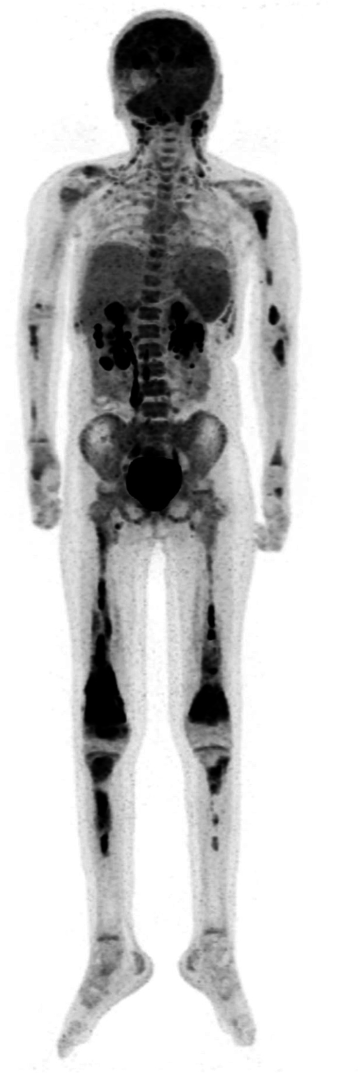
Figure 2.

18F-FDG PET scan on post-tisagenlecleucel relapse. 18F-FDG PET illustrates sacral sparing, yet, grossly patchy enumerable osseous and lymphoid lesions (in context of flow cytometry MRD from bone marrow aspirate of <0.01%, with detectable flow cytometry MRD in peripheral blood of 0.06%). 18F-FDG PET, 18F-fluorodeoxyglucose positron emission tomography; MRD, minimal residual disease.
