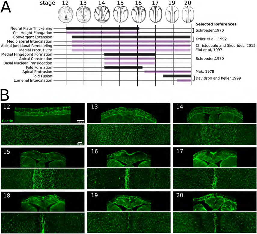
Figure 1: Concurrent mechanical processes shape the neural tube in Xenopus laevis.
A) Stage-dependency of specific tissue deforming processes (black bars) and cell behaviors (purple bars) that accompany the different phases of neurulation in Xenopus laevis. We refer interested readers to a similar diagram describing tissue movements and cell behaviors during stages of chick neurulation (Schoenwolf and Smith, 1990). B) Transverse sections and maximally z-projected en face sections of F-actin stained cell outlines of the posterior neural and non-neural dorsal ectoderm in fixed Xenopus laevis embryos showing cell and tissue morphological changes at each stage of neurulation.

