
Fig. 3.
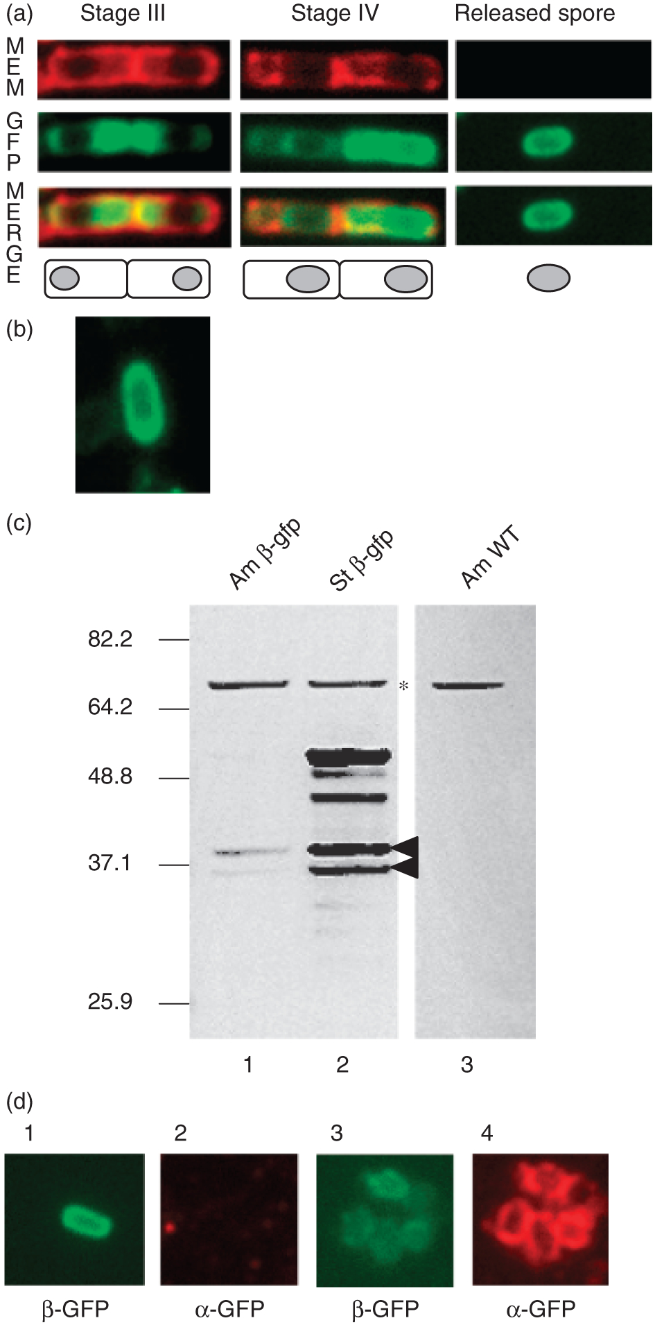
Fluorescence microscopic and Western blot analysis of Cotβ-GFP in Ames and Sterne strain spores and sporulating cells. (a) Fluorescence microscopic localization of Cotβ-GFP in strain RG134. Cells were grown at room temperature and analyzed 24 h (corresponding to stage III), 28 h (corresponding to stage IV), and 48 h (after spore release) after inoculation. Membrane staining (MEM) is shown in the upper panels, GFP fluorescence (GFP) is shown in the middle panels, and the merged (MERGE) image is shown in the lower panels. Forespore membranes are not stained due to the exclusion of the dye by the coat. The sporangia pictured in the merged images are cartooned below for clarity; spores and forespores are indicated by dark grey ovals, and mother cells are indicated by white rounded rectangles. Released spores in (a) were prepared by water washing. Fluorescence of Hypaque-purified released spores was indistinguishable from that of water-washed spores (data not shown). (b) Deconvolved micrograph from a spore from strain MGM37. (c) Spore coat extracts of cotβ-gfp fusion bearing spores, from strains Ames-JAB-10 and RG134 (lanes 1 and 2, respectively), or from the wild-type Ames strain spores (lane 3) were fractionated using SDS-PAGE and transferred to a PVDF membrane, and then probed with anti-GFP antibodies. The asterisk (*) indicates a cross-reacting species, and arrowheads indicate the Cotβ-GFP-specific doublet that is present in both fusion-bearing strains. (d) After sporulation for 2 days at room temperature, spores bearing cotβ-gfp (from strain RG134, panels 1 and 2), or bearing cotβ-gfp and a cotE mutation (from strain MGM37, panels 3 and 4) were collected and imaged by fluorescence (β-GFP) or IFM (α-GFP).