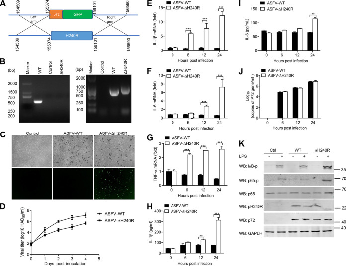
FIG 7.
ASFV-ΔH240R induces higher levels of mRNA transcription and secretion of inflammatory cytokines. (A) Schematic representation of the experimental design for generation of ASFV-ΔH240R. The H240R gene segment was replaced with the p72-EGFP reporter gene cassette. (B) Verification of the H240R gene deletion in ASFV-ΔH240R. The H240R gene was examined using PCR in both ASFV-WT (lane 1) and ASFV-ΔH240R (lane 3) using primers H240R-F/R, and the p72-EGFP gene was examined using PCR in both ASFV-WT (lane 5) and ASFV-ΔH240R (lane 6) using primers 72EGFP-F/R. Agarose gel (1%) showing the result of the PCR to amplify of the genomic segment containing the targeted gene. (C) Hemadsorption characteristics of ASFV-ΔH240R. PAMs were infected with ASFV-WT or ASFV-ΔH240R. At 24 hpi, the cells were observed by microscope. (D) Growth kinetics of ASFV-ΔH240R and ASFV-WT in PAMs. PAMs were infected with ASFV-ΔH240R or ASFV-WT (MOI, 0.01), and samples were taken from three independent experiments at the indicated time points and titrated. (E to I) Analysis of inflammatory cytokine expression and secretion in PAMs infected with ASFV-WT or ASFV-ΔH240R; PAMs were infected with ASFV or ASFV-ΔH240R at the same number of genome copies (108 for 0, 6, 12, and 24 h. The mRNA levels of Il-1β) (E), Il-6 (F), and Tnf-α (G) in PAMs were detected by qPCR, and the secreted IL-1β (H) and IL-6 (I) in cell supernatants were detected by ELISA. (J) The genomic copy numbers of ASFV were detected by qPCR. (K) Immunoblot analysis of the degradation and phosphorylation of IκB and p65 in PAMs upon ASFV-WT or ASFV-ΔH240R infection. PAMs were mock-infected or infected with ASFV-WT or ASFV-ΔH240R and then mock-treated or treated with LPS for 6 h. The expression of IκB, p65, pH240R, and GAPDH and the phosphorylation of IκB and p65 were detected by Western blotting. All assays were independently repeated at least three times. The data are shown as the mean ± SD; n = 3. **, P < 0.01; ***, P < 0.001.

