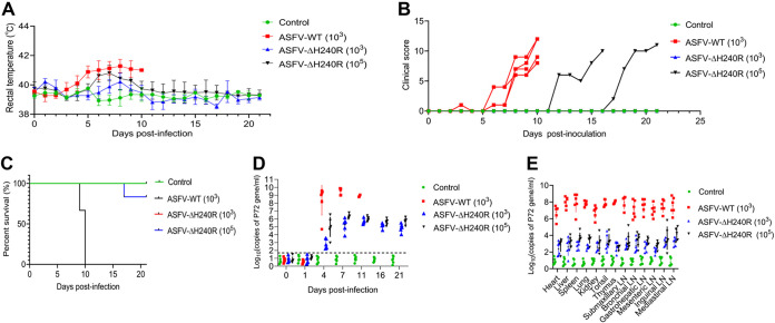
FIG 8.
Deletion of the H240R gene reduces ASFV pathogenicity in pigs. (A to C) The rectal temperature measurements (A), clinical score (B), and survival rates (C) of ASFV-WT- and ASFV-ΔH240R-challenged pigs. The different groups of pigs were unchallenged (control, n = 4), challenged with 103 HAD50 of parental ASFV-WT (n = 6), 103 HAD50 of ASFV-ΔH240R (n = 6), or 105 HAD50 of ASFV-ΔH240R (n = 6). The rectal temperature measurements (A), clinical scores (B), and survival rates (C) were recorded daily until day 21 postchallenge. (D) ASFV DNA copy numbers in blood samples from ASFV-WT- and ASFV-ΔH240R-challenged pigs. The blood samples were collected at 0, 1, 4, 7, 11, 16, and 21 dpi from unchallenged or ASFV-WT-challenged (103 HAD50) or ASFV-ΔH240R-challenged (103 and 105 HAD50) pigs to detect the viral DNA copy number by qPCR. (E) ASFV DNA copy number in different tissues from ASFV-WT- and ASFV-ΔH240R-challenged pigs. At 21 dpi, the surviving pigs were euthanized, and tissues including the heart, liver, lung, kidney, spleen, tonsil, thymus, and six lymph nodes (inguinal lymph node, submaxillary lymph node, bronchial lymph node, mesenteric lymph node, mediastinal lymph node, and gastrohepatic lymph node) were collected from all pigs for viral DNA quantification by qPCR. For pigs that died before 21 dpi, the indicated tissue samples were collected at the time of death.

