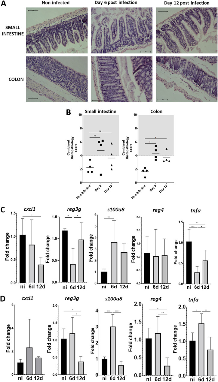
FIG 3.
K. pneumoniae colonization does not produce severe tissue damage. (A) Hematoxylin-eosin staining of tissue at different days postinfection with Kp52145. (B) Quantification of the histopathology changes upon infection. Each dot represents a different mouse. Five slides per section of tissue, per time point and per mouse, were scored. (C and D) The cxcl1, reg3g, s100o8, reg4, and tnfa mRNA levels were assessed by qPCR in the small intestine (C) and in the colon (D) of noninfected mice (black bars) and infected mice (gray bars) at 6 (6 d) and 12 (12 d) days. 5 to 7 mice were analyzed in each group. In panel A, the images are representative of four infected mice. Each value is presented as the mean ± SD. *, P ≤ 0.05; **, P ≤ 0.01; ***, P ≤ 0.001; ****, P ≤ 0.0001 for the indicated comparisons, which were determined using a one way-ANOVA with the Bonferroni correction for testing multiple comparisons. No other comparison is significant (P > 0.05).

