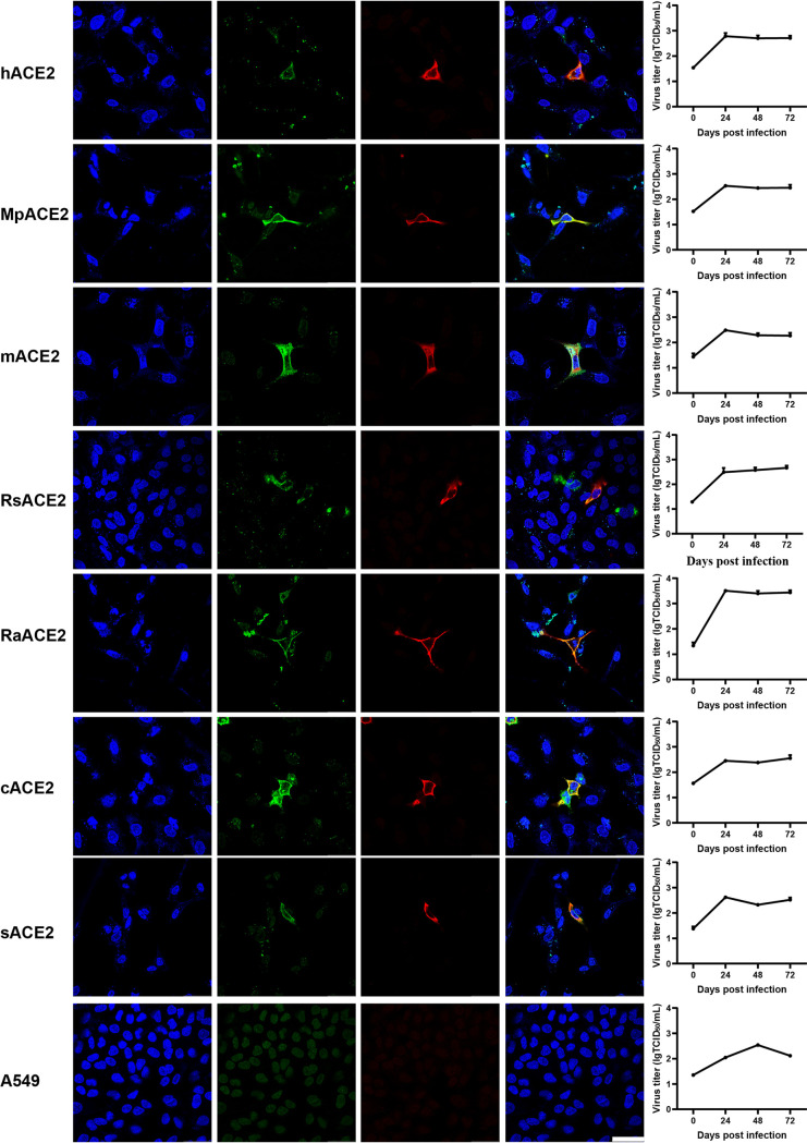
FIG 1.
Analysis of the ACE2 usage spectrum of MpCoV-GX using immunofluorescence assay and RT-qPCR. A549 cells with and without expression of ACE2 from different species were infected with MpCoV-GX at an MOI of 1.0. ACE2 expression was detected using a mouse anti-S tag monoclonal antibody and an FITC-labeled goat anti-mouse IgG(H+L). At 24 hpi, virus replication was detected using rabbit serum against the SARSr-CoV-Rp3 Np and a Cy3-conjugated goat anti-rabbit IgG. Nuclei were stained with DAPI. hACE2, human ACE2; MpACE2, Manis pentadactyla ACE2; mACE2, mouse ACE2; RsACE2, Rhinolophus sinicus ACE2; RaACE2, Rhinolophus affinis ACE2; sACE2, swine ACE2; cACE2, civet ACE2; A549, no ACE2 expression. The columns (from left to right) show staining of nuclei (blue), ACE2 expression (green), virus replication (red), merged triple-stained images, and RT-qPCR results, respectively (n = 3). Error bars represent the standard error. Staining patterns were examined using a confocal microscope. Bars, 40 μm.

