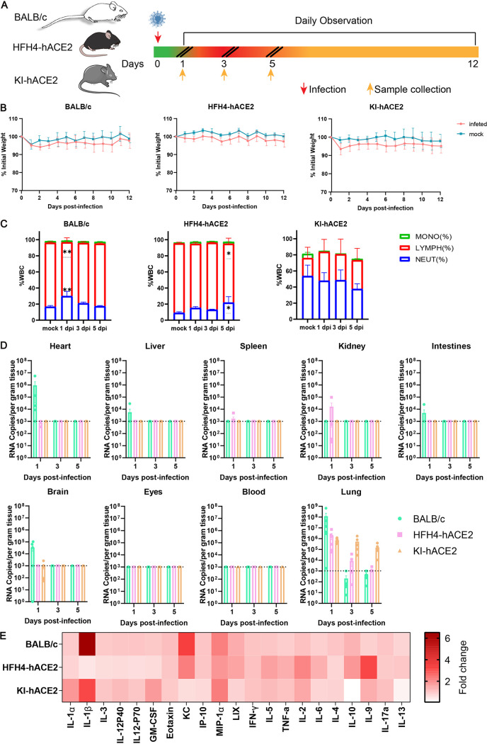
FIG 4.
Clinical symptoms, viral replication, and tissue distribution of MpCoV-GX. (A) Mice were mock infected (n = 4) or intranasally infected (n = 8) with 4 × 10⁵ TCID50 of MpCoV-GX and sacrificed for tissue and blood sampling at 1, 3, and 5 dpi. (B) Mouse body weight was monitored until 12 dpi. (C) Peripheral blood was collected from mock-infected (n = 6) and virus-infected mice at 1 (n = 6), 3 (n = 6), and 5 (n = 6) dpi. The white blood cell (WBC) population was measured using a ProCyte Dx hematology analyzer. Error bars indicate the standard error. Statistical significance was measured by two-way analysis of variance in comparison with the mock infection group. **, P < 0.01; ***, P < 0.001; ****, P < 0.0001. (D) Mice (n = 6) were sacrificed and tissue (heart, liver, spleen, lung, kidney, intestine, brain, and eyes) and blood samples were collected at 1, 3, and 5 dpi, respectively, and viral RNA copy numbers were determined using RT-qPCR. (E) Serum cytokine/chemokine heatmap in infected mice at 1 dpi. GM-CSF, granulocyte-macrophage colony-stimulating factor; IFN-γ, gamma interferon; TNF-a, tumor necrosis factor alpha.

