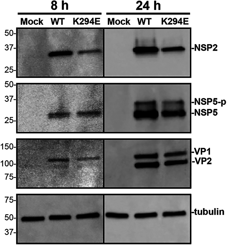
FIG 7.

Western blot of NSP2 and NSP5. MA104 cells were either mock infected or infected with rRV-WT or rRV-NSP2K294E at an MOI of 3 PFU per cell for 8 or 24 h. Lysates were subjected to Western blotting using αNSP2, αNSP5 αVP1/VP2, or αtubulin as a loading control. Secondary antibodies conjugated to HRP were used for detection. Molecular weight markers (in kDa) are shown to the left of the blots, and the locations of the protein of interest are labeled on the right. Hyper-phosphorylated NSP5 (NSP5-p).
