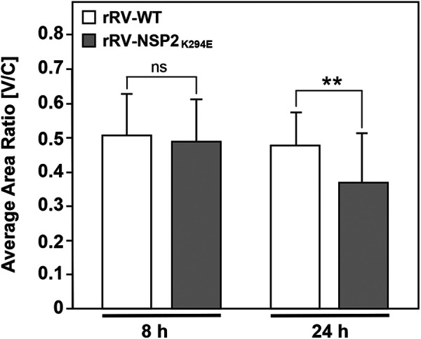
FIG 9.

Perinuclear localization of viroplasms. MA104 cells on glass coverslips were infected with rRV-WT or rRV-NSP2K294E at an MOI of 3 PFU per cell for 8 or 24 h. Cells were fixed with methanol and stained using αNSP2 and an Alexa-546-conjugated secondary antibody. Nuclei were stained using Hoechst. Confocal microscopy was used to determine the localization of NSP2 (561 nm) and nuclei (405 nm). Perinuclear condensation (V/C ratio) was determined as described in Materials and Methods. Statistical significance was determined by a two-tailed, unpaired t test. ns, not significant. Asterisks (**) indicate a P value of <0.01.
