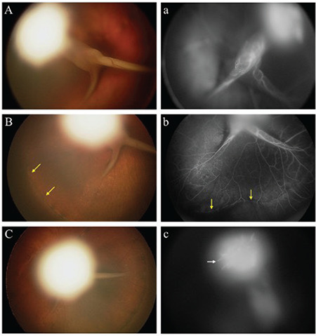
Figure 11.

Persistent fetal vasculature in a 6-month-old boy presenting with leukocoria and exotropia of the right eye.40 Color fundus photograph of the right eye revealed a fibrovascular stalk extending from the optic nerve to the posterior lens capsule (A, B, C). Fluorescein angiography of the right eye shows retinal vessels in posterior retinal folds pulled anteriorly into the stalk up to the posterior lens (a, c), as well as peripheral retinal capillary nonperfusion (b). Reprinted from Pictures & Perspectives, 124/4, Jeng-Miller KW, Joseph A, Baumal CR, Fluorescein Angiography in Persistent Fetal Vasculature, Page 455, Copyright (2016), with permission from Elsevier
