
Figure 9.
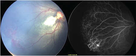
A case of Coats’ disease with a peripheral avascular retina, mimicking familial exudative vitreoretinopathy. Color fundus photograph of Coats’ disease shows peripheral vascular proliferations and branched vessels, and exudative changes in the posterior pole. Fluorescein angiography of the same case shows peripheral avascular retina with bulb-shaped telangiectasias (white arrow), suggesting the diagnosis of Coats’ disease (Courtesy of Dr. Şengül Özdek)