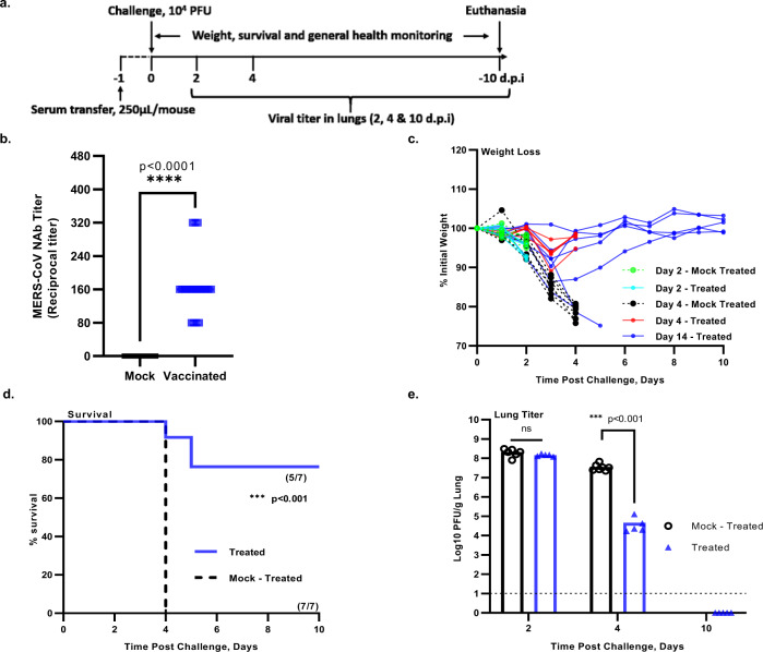
Fig. 7. Challenge of hDPP4 KI mice after passive transfer of immune sera from mice immunized with the DUB-negative rMERS-CoVMA.
a Timeline of immunization, serum transfer, challenge and clinical outcomes. Naive hDPP4 KI mice were injected intraperitoneally with 0.25 mL of pooled serum from hDPP4 KI mice immunized with a single shot of the rMERS-CoVMA-DUBneg. At 24 h post serum transfer, mice were intranasally challenged with a lethal dose of 104 PFU of rMERS-CoVMA. b Neutralizing activity of the pooled immune sera from hDPP4 KI mice immunized with rMERS-CoVMA-DUBneg (pooled mock-vaccinated: n = 28; pooled rMERS-CoVMA-DUBneg: n = 20). Black horizontal lines indicate mean reciprocal titers and squares (symbols) indicate individual values per group. An unpaired two-tailed t test was used to determine significant differences between the pooled mock-vaccinated neutralizing titers (black squares) and the pooled DUB-negative rMERS-CoVMA vaccinated neutralizing titers (shown in blue squares), ****P < 0.0001. c Body weight kinetics after virus challenge and d survival (n = 7) were monitored daily for 10 days. e On days 2, 4, and 10 post challenge, lung tissues were collected for virus titration by plaque assay on Huh7 cells. The mean ± SEM per group and the virus titer in PFU per gram of lung tissue are presented. Symbols represent individual mice. The limit of detection for infectious viral progeny is 10 PFU/g lung and is indicated with a dashed line. Statistical comparisons between means were performed by Student’s t test (unpaired tqo-tailed): ***P < 0.001. Source data are provided as a Source Data file.

