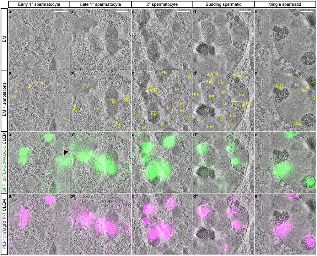
Fig. 6 ∣. PEI granules are associated with membranous organelles.
a-e, Representative dual-color CLEM images acquired in indicated germ cells of spermatogenesis (indicated at the top) of high-pressure frozen adult males expressing GFP::3xFLAG::WAGO-3 and PEI-1::mTagRFP-T. The four rows show the EM-only (a-e), EM with annotations (a’-e’), EM with GFP::3xFLAG::WAGO-3 fluorescence (a”-e”), and EM with PEI-1::mTagRFP-T fluorescence (a’”-e’”). The GFP::3xFLAG::WAGO-3-only positive focus in panel a” (black arrow head) is adjacent to the nucleus, and most likely representing a P granule. Images represent two biologically independent experiments. Annotations: EM - electron microscopy, CLEM - correlative light and electron microscopy, N – nucleus, M – mitochondrion, MO – membranous organelle, FB – fibrous body, GO – Golgi complex, NH – peri-nuclear halo, D – DNA. Scale bars: 500 nm. Source data are provided.

