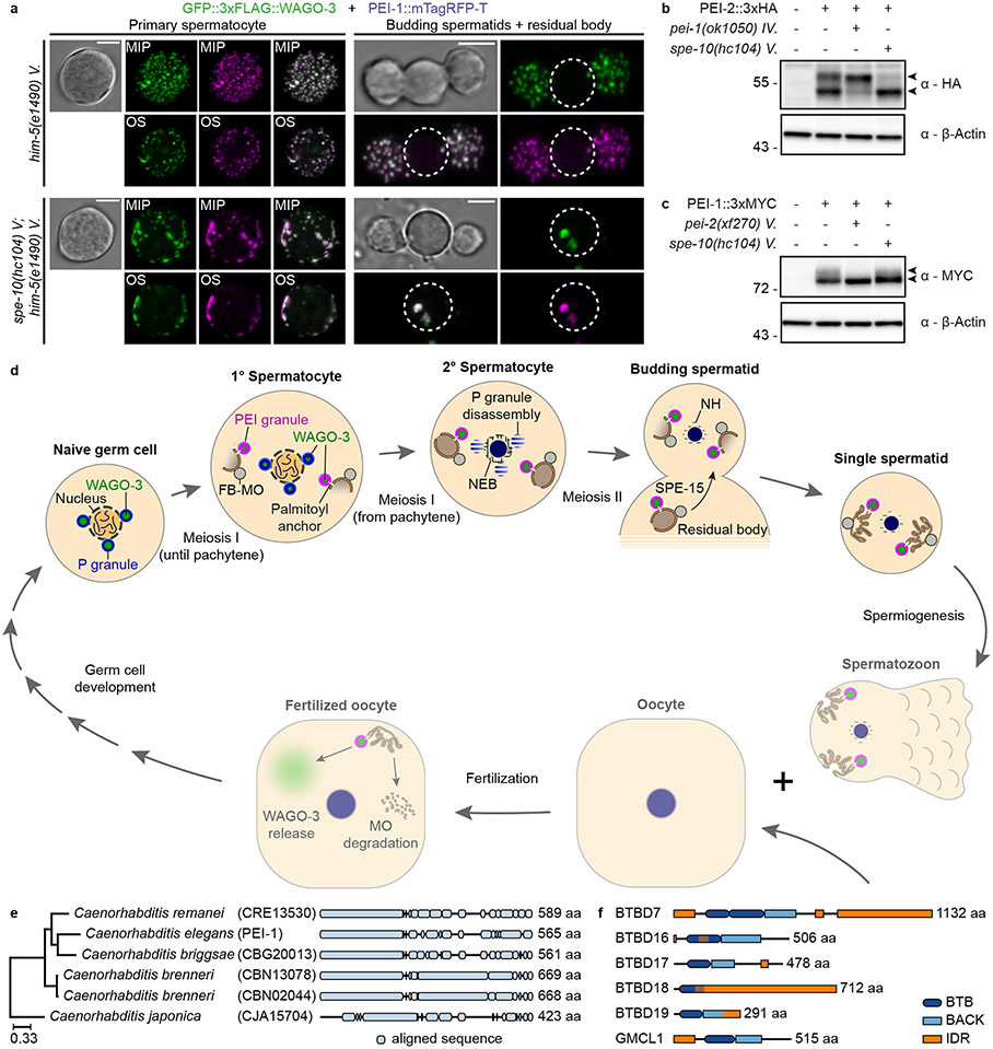
Fig. 8 ∣. PEI granule formation and segregation depends on S-palmitoylation.
a, Confocal maximum intensity projections and optical sections of isolated, male-derived spermatocytes and budding spermatids expressing GFP::3xFLAG::WAGO-3 and PEI-1::mTagRFP-T in indicated mutants. Strains contained him-mutations to increase the frequency of males in the cultures. Dashed circles indicate residual bodies. Images represent two biologically independent experiments. MIP - maximum intensity projection. OS – optical section. Scale bars: 4 μm. b-c, Whole-worm extracts of late-L4 stage hermaphrodites were separated via SDS-PAGE, followed by Western transfer and chemiluminescence detection of PEI-2::3xHA (b) and PEI-1::3xMYC (c) in indicated mutants. The doublet signals of both proteins are indicated by arrow heads. β-actin served as loading control. Unprocessed original scans of blots are provided in source data. The experiment in b and c has been performed once. d, We provide data for the top half of the model: WAGO-3 starts in P granules, gradually moves to PEI granules that associate with FB-MOs via S-palmitoylation, which ensures spermatid localization via SPE-15 dependent transport. The bottom half is hypothetical, and depicted in reduced opacity. We speculate that PEI granules release their content into the oocyte, helping to establish/maintain silencing of specific targets (see Extended Data Figure 1). Schematic representation is not to scale. Annotations: MO – membranous organelle, FB – fibrous body, NH – peri-nuclear halo. e, Phylogenetic analysis showing PEI-1 conservation within the Caenorhabditis genus. The phylogenetic three was generated using EggNOG (v4.5.1). PEI-1 was defined as query and compared to all eukaryote entries. f, Protein length and domain composition of six human BTB domain-containing proteins that resemble PEI-1 protein composition.

