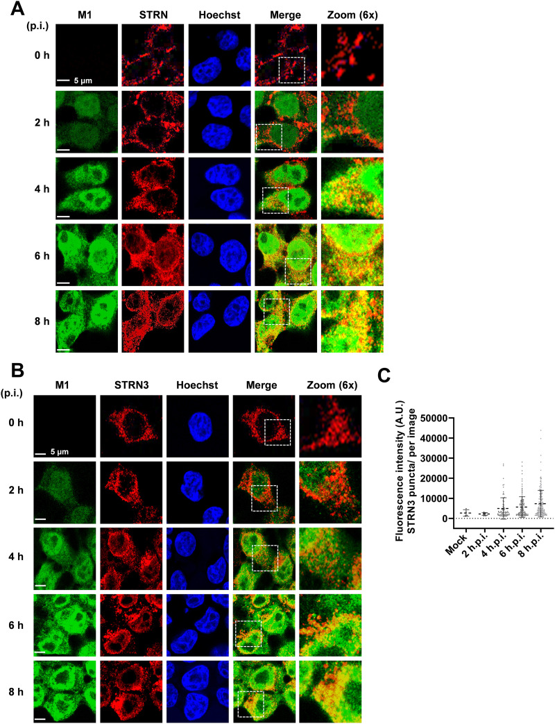
FIG 7.
IAV-mediated relocalization of STRN and STRN3. (A) 293T cells were infected with SC35M (MOI = 2) for various periods as shown. STRN and the viral protein M1 were detected by indirect immunofluorescence, and nuclei were revealed by staining of nuclear DNA with Hoechst dye. Representative confocal images from two independent experiments are shown; yellow areas indicate colocalization, and the boxed areas are displayed in 6× magnification. Representative confocal images from two independent experiments show intracellular colocalization between STRN and the viral M1 protein. (B) The same experiment was performed as described for panel A, with the exception that M1 was costained with STRN3. (C) Quantification of the number of STRN3 puncta per image. Data are means ± SD from one representative experiment where at least 150 cells per condition were quantified. A.U., arbitrary units.

