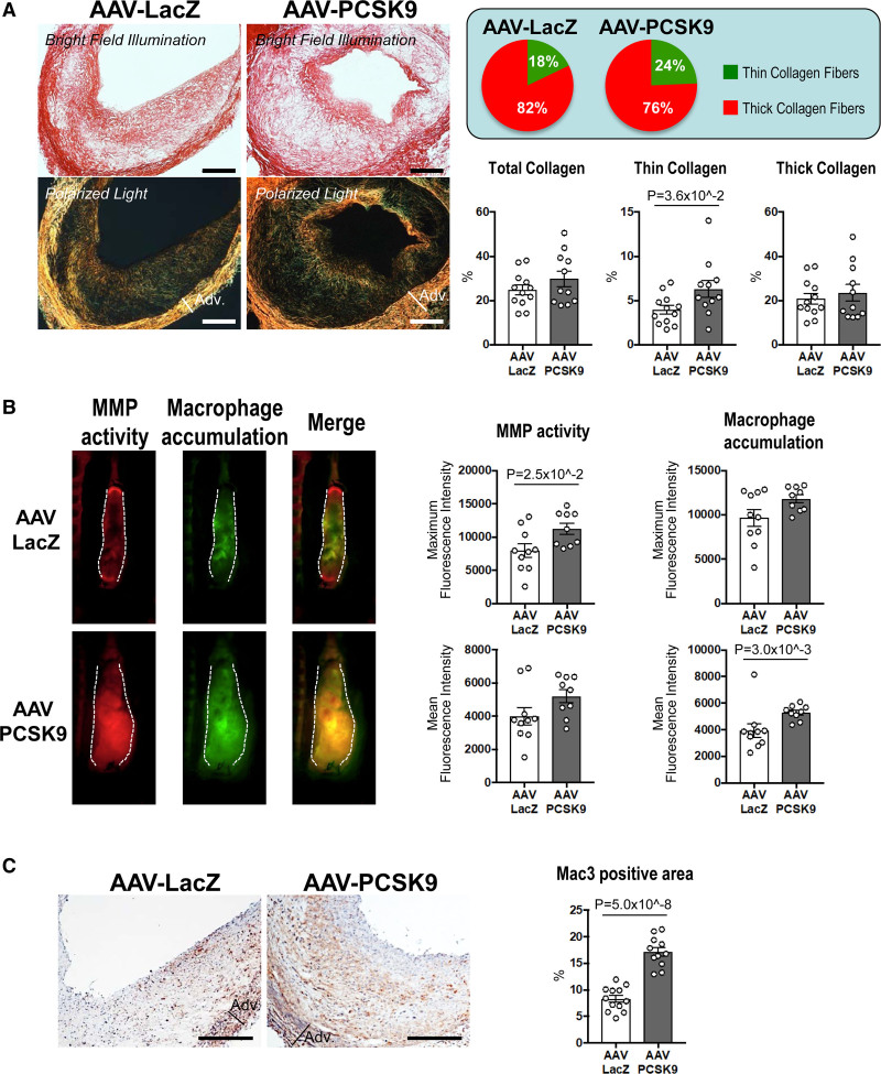
Figure 2.
The effects of Adeno-associated virus (AAV)-PCSK9 (proprotein convertase subtilisin/kexin 9) on collagen thinning and macrophage accumulation in vein graft lesions of Ldlr-/- mice. A, Picrosirius red staining of vein grafts without (top) or with (bottom) polarized light (n=12 and 11 for AAV-LacZ and AAV-PCSK9 group, respectively). Scale bars indicate 500 μm. Circle graphs show the percentage of thin (green) and thick (red) collagen fibers compared with total fibers. B, Intravital microscopy images of MMPSense 680 (red) and AminoSPARK750 (green) in vein grafts for visualization of MMP activity and macrophage accumulation, respectively (n=10 and 9 for AAV-LacZ and AAV-PCSK9 group, respectively). C, Mac3 (macrophages) staining in vein grafts. Scale bars indicate 500 μm. P value was calculated by Mann-Whitney U-test (thick collagen in A, macrophage accumulation mean fluorescence intensity in B) and unpaired Student t test (C). Data are reported as mean ± SEM.

