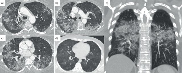
FIGURE 1.
High-resolution computed tomography (CT) images of the chest from patients with diffuse alveolar haemorrhage (DAH) in pulmonary renal syndrome. a–c) The axial CT slices show extensive bilateral mixed consolidative and ground-glass opacities with a mid to lower zone predominance admixed with coarsened interlobular septa, appearances typical for DAH. d) Milder disease is shown on an axial CT slice with patchy ground-glass opacities bilaterally. e) A coronal CT chest image highlights the mid to lower zone predominance in DAH.

