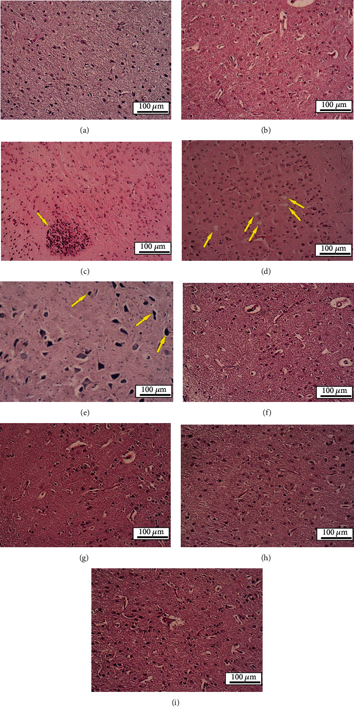
Figure 10.

Histological sections of experimental groups' brain. H&E staining, ×100. (a, b) Normal saline and GSH-PMAA group, respectively: histological structure was normal. (c) GSH-PMAA-EDV 250 μg/kg group: microglial nodule in the brain was observed (arrow). (d, e) TGI group: vacuolation and degeneration (arrows in (d)) and basophilic neuronal necrosis (arrows in (e)) were observed. (f–i) Received GSH-PMAA-EDV 5, 20, 40, and 250 μg/kg groups, respectively: histological structure was normal.
