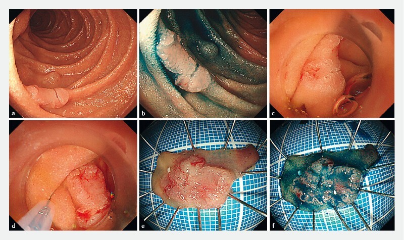
Fig. 1.

Example of gel immersion endoscopic resection of a superficial nonampullary duodenal epithelial tumor. a–d Endoscopic views showing: a,b a superficial 11-mm elevated and depressed lesion in the second part of the duodenum; c the favorable visual field after the lesion had been immersed in a gelatinous liquid; d a sufficient horizontal margin being easily confirmed with the lesion under the gelatinous liquid. e,f Macroscopic appearance of the en bloc resected lesion after it had been easily captured using an electrocautery snare, resulting in a specimen of 25 × 10 mm, containing a tumor of 11 × 8 mm that was pathologically diagnosed as a well-differentiated adenocarcinoma with negative margins.
