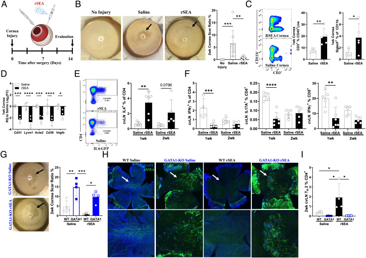
Fig. 4.
rSEA immunotherapy promotes repair in corneal injury. (A) Corneal debridement injury model timeline. (B) Corneal gross images and scar ratio assessment at 2 wk after injury treated with saline vehicle or rSEA, two study replications combined. (C) Flow cytometry populations isolated from the cornea 1 wk postinjury and treatment with saline or rSEA. (D) Corneal expression of genes associated with scar vascularization at 1 wk after injury and treatment with saline or rSEA. (E) Representative flow plots of 2 wk after injury 4get cervical LNs (cvLN) and % TH2 populations at 1 wk vs. 2 wk after injury and treatment with saline or rSEA. (F) ICS from cvLN of TH1, TH17, and IFNγ+CD8+ T cells (%) at 1 and 2 wk after injury and treatment with saline or rSEA. (G) Representative gross images of wounded corneas from GATA1 KO mice treated 2 wk with saline or rSEA and scar area ratio assessment. (H) Immunofluorescent staining of nuclei (DAPI, blue) and α-SMA (green) on corneas 2 wk postinjury and treatment with saline or rSEA in WT vs. GATA1 KO mice. (I) cvLN ICS for IL-4 in WT vs. GATA1 KO mice 2 wk postinjury and treatment with saline or rSEA. Statistical tests represent all biological replicates, and all experiments were replicated at least twice. Graphs show mean ± SD (B–F, G, and I), n = 4 to 6. *P < 0.05, **P < 0.01, ***P < 0.001, and ****P < 0.0001 by one-way ANOVA with Tukey’s multiple comparisons (B) and two-way ANOVA with Sidak’s multiple comparisons (D–F, G, and I).

