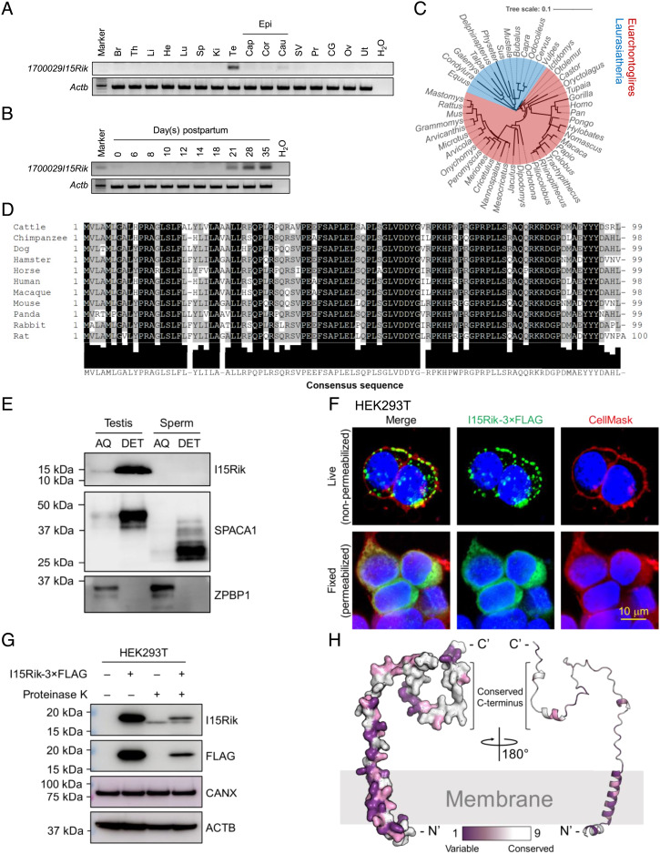
Fig. 1.
1700029I15Rik Is a Testis-Enriched Type-II Transmembrane Protein Conserved in Mammals and Expressed during Spermiogenesis. (A) Analysis of 1700029I15Rik expression in various mouse tissues by RT-PCR. Br, brain; Th, thymus; Li, liver; He, heart; Lu, lung; Sp, spleen; Ki, kidney; Te, testis; Cap, caput epididymis; Cor, corpus epididymis; Cau, cauda epididymis; Epi, epididymis; SV, seminal vesicle; Pr, prostate; CG, coagulating gland; Ov, ovary; Ut, uterus. The expression of β-actin (Actb) was analyzed as a loading control. (B) Analysis of 1700029I15Rik expression in mouse testes during postnatal development. (C) Phylogenetic tree depicting the evolutionary conservation of 1700029I15Rik in mammals. The tree was visualized using the interactive Tree of Life (iTOL) (16). Red and blue highlighted species belong to Euarchontoglires and Laurasiatheria, respectively. (D) Multiple sequence alignment of 1700029I15Rik orthologous proteins in 11 mammalian species. The Lower panel indicates the consensus sequence and the extent of amino acid conservation. (E) Western blot detection of 1700029I15Rik (I15Rik) in mouse testes and sperm fractionated by Triton X-114. SPACA1 and zona pellucida binding protein 2 (ZPBP1) were analyzed as positive controls for the proteins enriched in the AQ and DET phases, respectively. (F) In vitro topological analysis of 1700029I15Rik by live cell immunostaining. HEK293T cells were transiently transfected with a plasmid encoding C-terminal 3 × FLAG-tagged 1700029I15Rik. Live or fixed HEK293T cells were probed with an anti-FLAG antibody and an Alexa Fluor™ 488-conjugated secondary antibody. Cell membranes and nuclei were visualized by CellMask™ deep red plasma membrane stain and Hoechst 33342, respectively. (G) In vitro proteinase K protection assay depicting the topology of 1700029I15Rik. Live HEK293T cells transiently expressing 1700029I15Rik-3 × FLAG were treated with proteinase K and subjected to protein extraction. The levels of 1700029I15Rik-3 × FLAG before and after the enzyme treatment were analyzed by Western blotting. CANX and ACTB were analyzed in parallel as loading controls. (H) 1700029I15Rik protein structure predicted by AlphaFold (AF-Q8CF31-F1) (17). The degree of residue conservation was determined by ConSurf (18) and plotted to the three-dimensional structures, with purple representing variable and white representing conserved.

