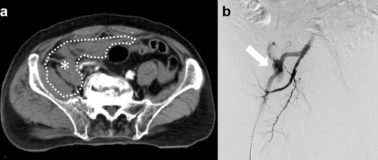
Fig. 3.
Pelvic contrast-enhanced CT images obtained during complete REBOA. a Delayed-phase (second phase) contrast-enhanced CT image shows a massive hematoma (asterisk) without extravasation of the contrast material. b Digital subtraction angiography of the right internal iliac artery obtained during REBOA balloon deflation shows massive extravasation from the iliolumbar artery, which represents “hidden” bleeding on contrast-enhanced CT during complete REBOA (arrow). We embolized the iliolumbar artery with NBCA. CT computed tomography; REBOA resuscitative endovascular balloon occlusion of the aorta; NBCA n-butyl cyanoacrylate

