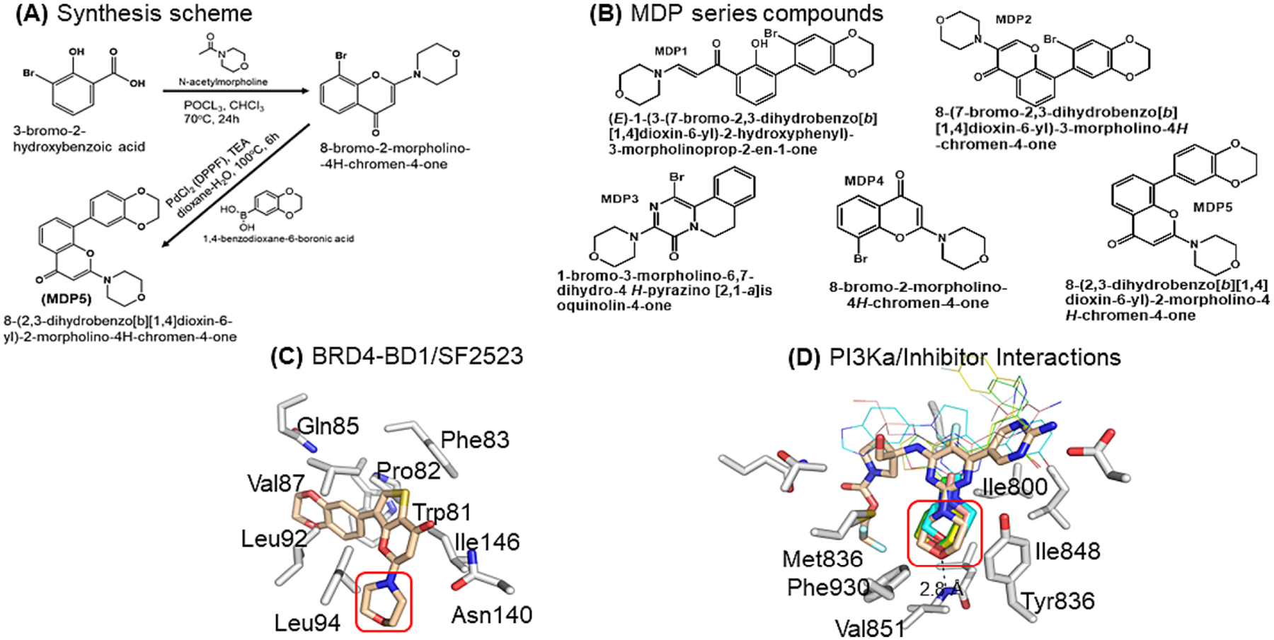
Fig. 1.

Inspirational structures for dual inhibitor of BRD-BDs and PI3Ka. A Synthesis scheme of MDP5. B Chemical structures of MDP series compounds. C X-ray crystal structure of BRD2-BD1/SF2523 complex (pdb accession: 5u28) highlighting the benzodioxane and thienopyranone interactions with the acetyllysine binding site and the lack of interaction between BD1 and the morpholino moiety of SF2523 (red box). D Xray crystal structures of PI3Ka/g (gray carbons, 7k6m) complexed with various morpholino-containing inhibitors (tan, cyan, pink, yellow, and green carbons representing pdb accession codes 7k6m, 3apf, 5jhb, 5xgh, 5xgj, respectively). The morpholino moiety binds in a hydrophobic pocket formed by the indicated PI3Ka residues and forms a pivotal hydrogen bonded interaction with the backbone amide of Val851 (red box). The narrow bonds indicate structurally variable regions of the 5 inhibitors.
