
Fig. 7.
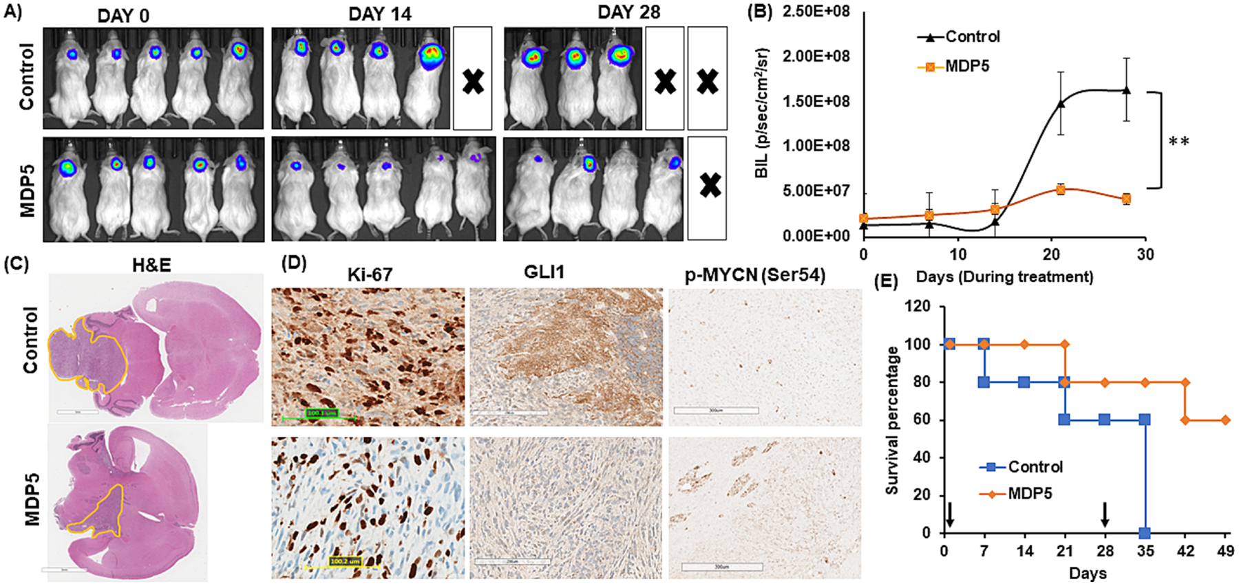
Antitumor efficacy of MDP5 after systemic administration into DAOY cell generated orthotopic MB bearing NSG mice. A Bioluminescence imaging of orthotopic MB bearing mice during the treatment. B Quantitation of BLI in MDP5 treated and control group (n = 3, **P < 0.01). C H&E staining of brain harvested from mice bearing orthotopic MB after treatment (Scale bar 3 mm). D IHC staining of tumor tissues for Ki-67, GLI1, and p-MYCN (Ser54) (Scale bar 100–300 μM). E Overall survival curve showing MDP5 treated mice survived significantly longer than the control group (n = 3).