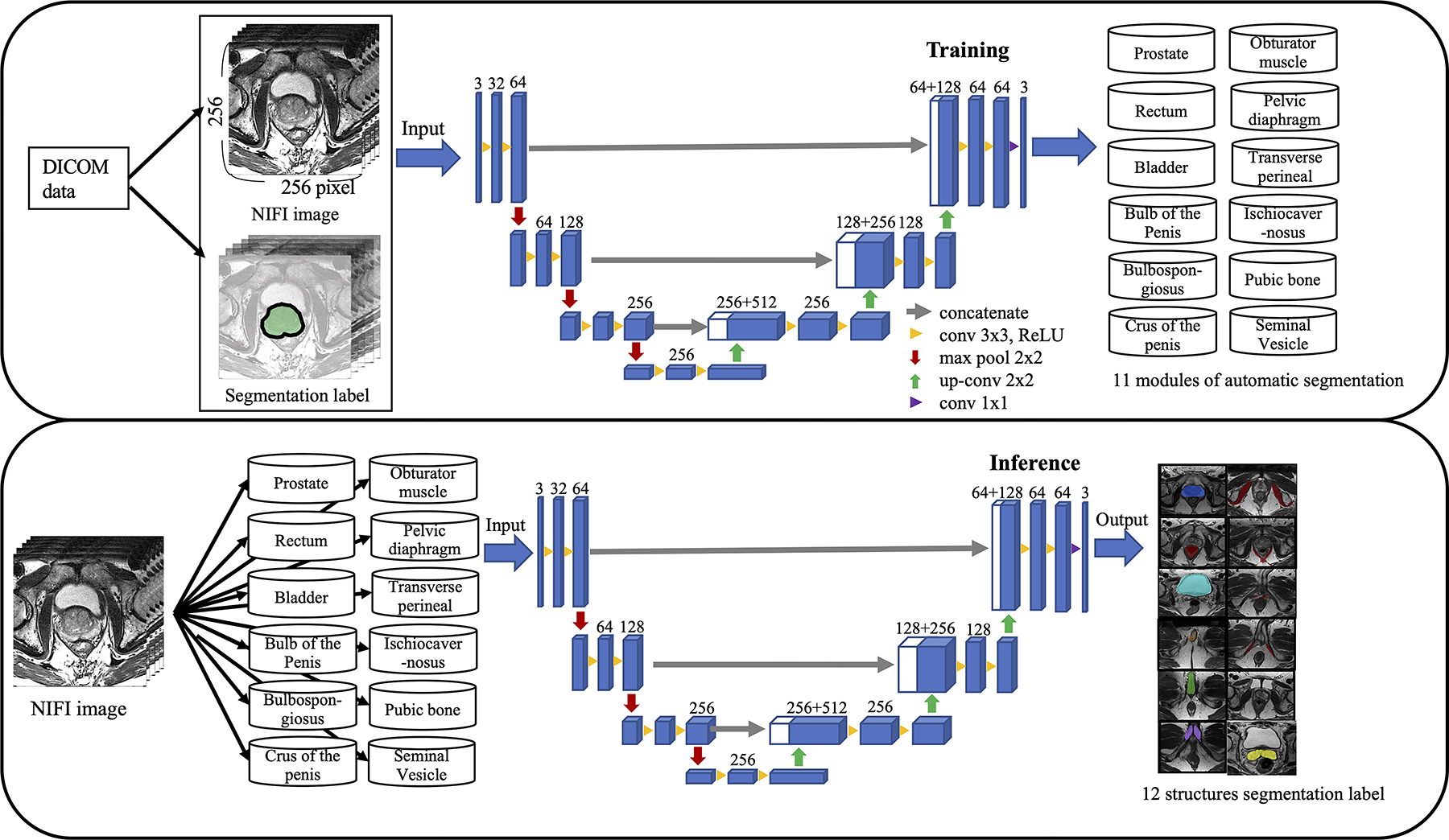
Fig. 1.

The overall framework of automatic segmentation for the prostate and extracapsular structures. Digital imaging and communications in medicine data were converted to the Neuroimaging Informatics Technology Initiative (NIfTI) images and manually segmented 12 structures. The training module was constructed by inputting the image data and label data of each of the 12 structures into the 3D U-Net as training data. Inference learned the label data by inputting the NIfTI image into the module of 12 structures
