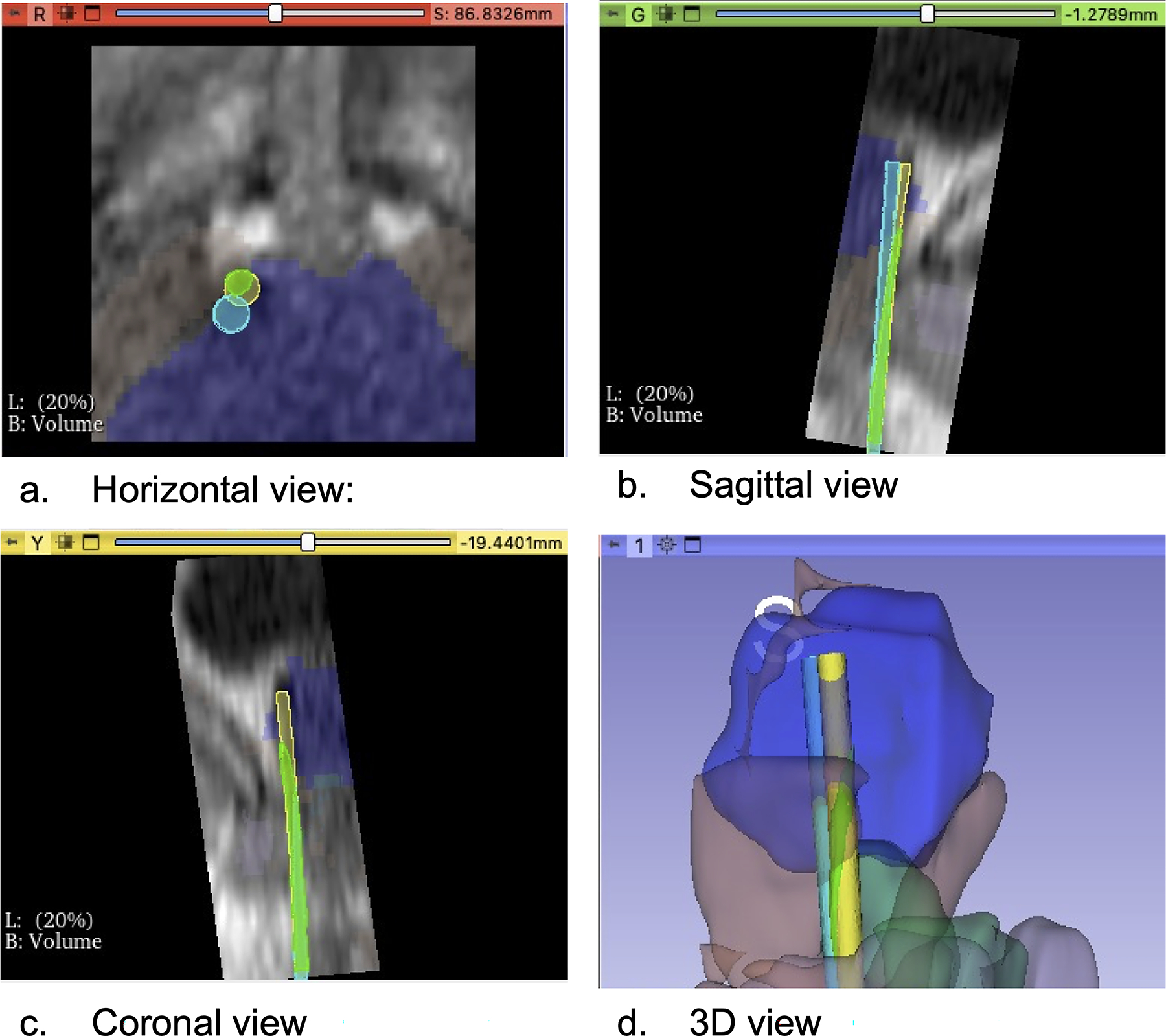
Fig. 6.

A segmentation label was automatically created using the MRI of case no.4, and needle deflection was predicted. a Horizontal view: The light blue circle represented the preoperatively planned path, the light green circle represented the needle path including prediction of needle deflection, and the yellow circle represented the deflected path through which the actual needle passed. b Sagittal view: The deflected path deviated from the planned path. c Coronal view: The predicted path overlapped with the deflected path, and we were able to predict the needle deflection. d 3D view: We visualized three paths in a 3D image: the blue structure was the prostate, the brown structure was the pelvic diaphragm muscle, the green structure was the bulb of the penis
