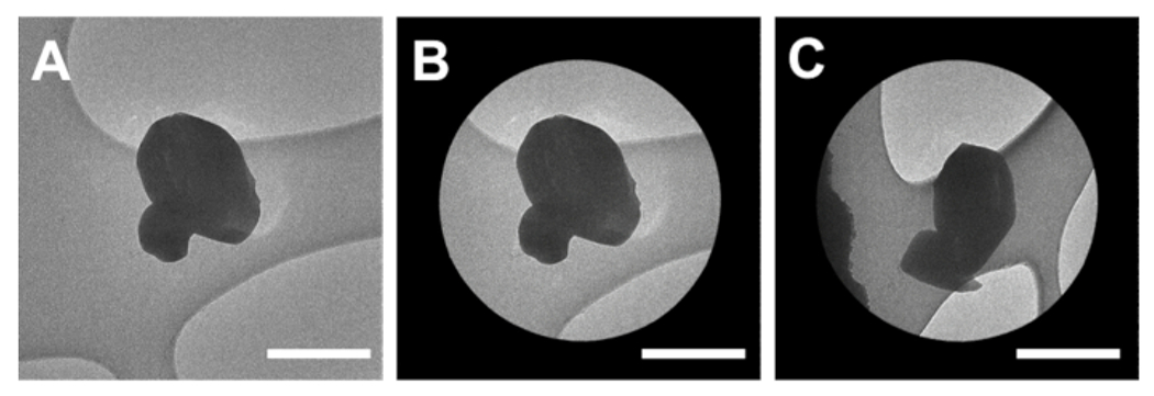
Figure 3: Screening and aligning microcrystals for MicroED data collection.

(A) High magnification micrograph of a microcrystal. (B) Micrograph of the isolated crystal within the selected area aperture. (C) The same microcrystal in the aperture with the stage tilted to −69 °. Scale bars all 1 μm. Please click here to view a larger version of this figure.
